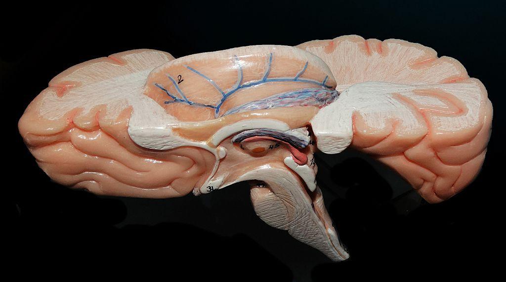
Scientists at the Sudha Gopalakrishnan Brain Centre (SGBC) of the Indian Institute of Technology, Madras, have created Anchor – the world’s most detailed 3‑D atlas of the human brainstem. By stitching together over 500 tissue sections from foetal, childhood and adult brains, the atlas maps more than 200 clusters of brain cells and nerve pathways with eight chemical markers.
Anchor bridges two worlds that have largely stayed separate: whole‑brain MRI, which shows the brain’s overall structure, and cellular pathology, which reveals it one cell at a time. The free online viewer lets users zoom from the entire brainstem down to individual neurons while preserving their exact spatial relationships.
Experts say the atlas could transform research into neurological diseases such as Alzheimer's, Parkinson’s, stroke, sudden infant death syndrome and even the long‑term effects of COVID‑19 on the brain. By comparing healthy and diseased brainstem maps, scientists hope to identify how disorders alter cell populations and pathways.
“It puts India on the international table,” says neuroscientist Shubha Tole of the Tata Institute of Fundamental Research. “We are seeing a visionary programme that integrates engineering, neuroscience and medicine.”
In future, SGBC plans to image more than 100 human brains at different life stages and disease states, creating a reference library that will reveal how diseases reshape the brain cell‑by‑cell.